


When the pulp, or inner nerve tissues, of your tooth become infected or damaged, a root canal is often the best treatment to restore the health and function of the tooth. Our dentist and team have received advanced training, and we use sedation techniques and anesthesia to ensure that your procedure is as comfortable as possible.
Root canal therapy is an endodontic treatment often preformed when injury or decay reaches deep into the tooth, beyond the outer layer of enamel, and infects the nerve tissues of your tooth. Root canal treatment can prevent your tooth from dying and requiring extraction.
During your root canal procedure, our dentist will perform the following steps:
- Examine and X-ray the tooth to determine that a root canal is the proper treatment.
- Numb the tooth and place a small protective sheet around it to prevent saliva or other particles from interfering with the procedure.
- Make an opening in the crown of the tooth.
- Use small instruments to reach into the pulp chamber and root canals. This removes damaged pulp and nerves, and shapes the inside of the canals for a filling.
- Fill the root canals with a filling and places a temporary cap or filling on the tooth’s crown to cover the opening.
- Replace the temporary restoration with a permanent one once it has been custom-made in a dental laboratory.
To help you gain a better understanding of what you can expect during your root canal treatment, we invite you to review the images and video below. Please call Smile Studio Dental at 703-982-2222 if you have any questions about receiving root canal therapy in Falls Church, Virginia, and to schedule your appointment with Dr. Eli Janabi or Dr. Zinah Shakir.
Endodontic Cases
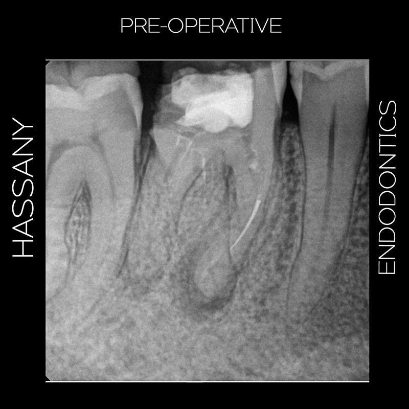
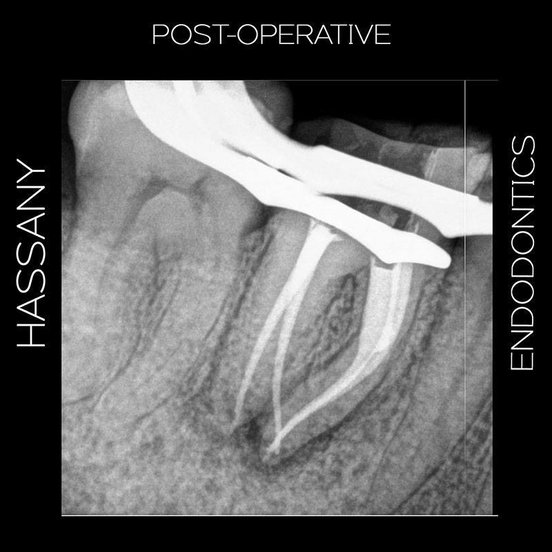
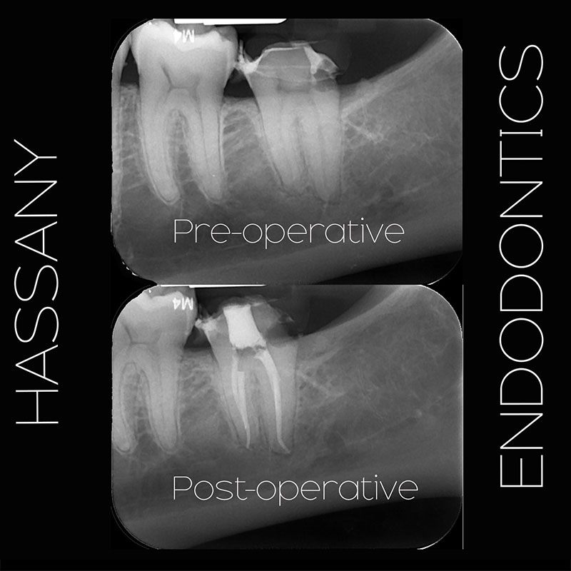
Case 1: Separated instrument
Patient referred by GP for separated instrument in the lower right first molar. Even with a microscope, negotiation was very difficult and done with hand instruments, rotary files and ultrasonic tips. After 30 minutes of negotiation and irrigation complete patency was obtained and preparation was completed at full working length with Protaper Next and Edge endo files. Obturation completed with standardized GP cones and back fill technique. Patient was referred back to GP for final restoration.

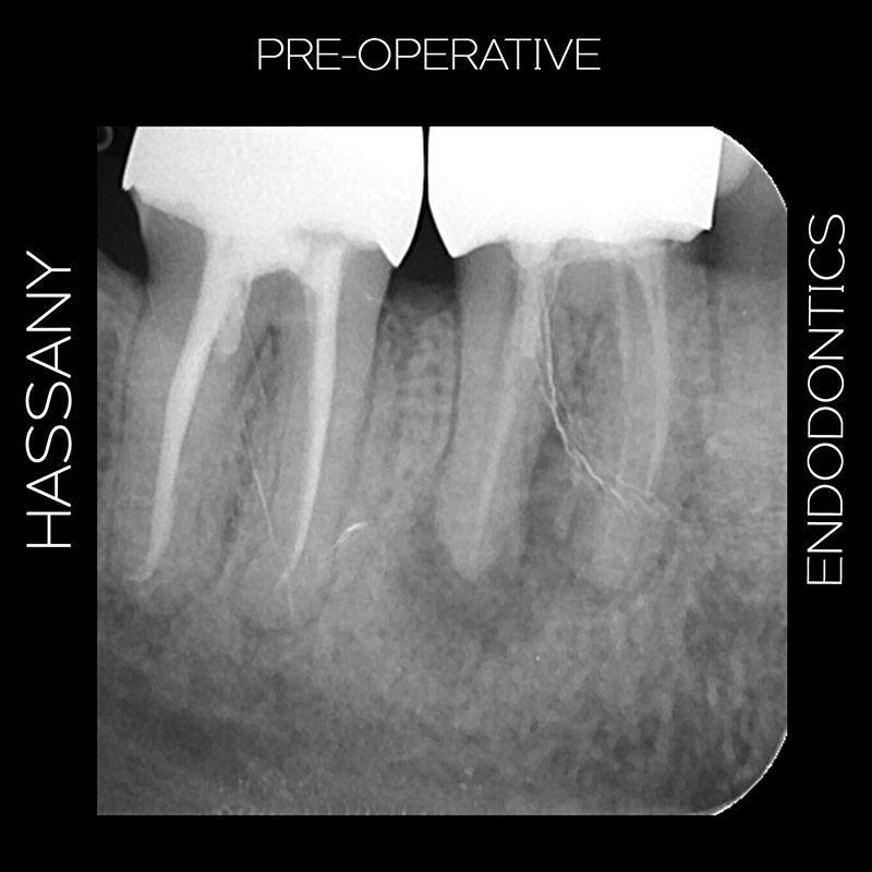
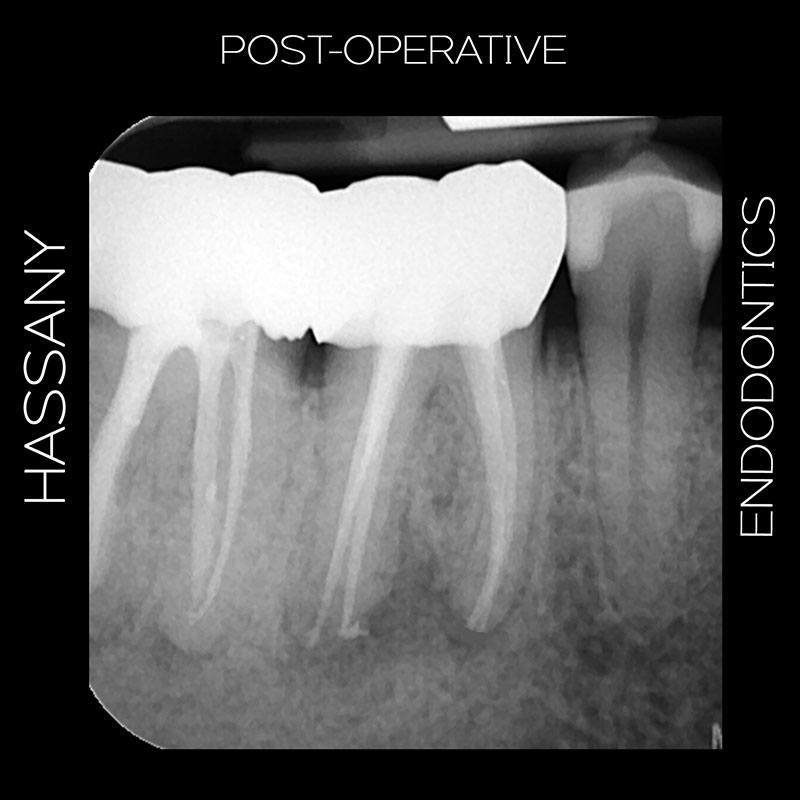
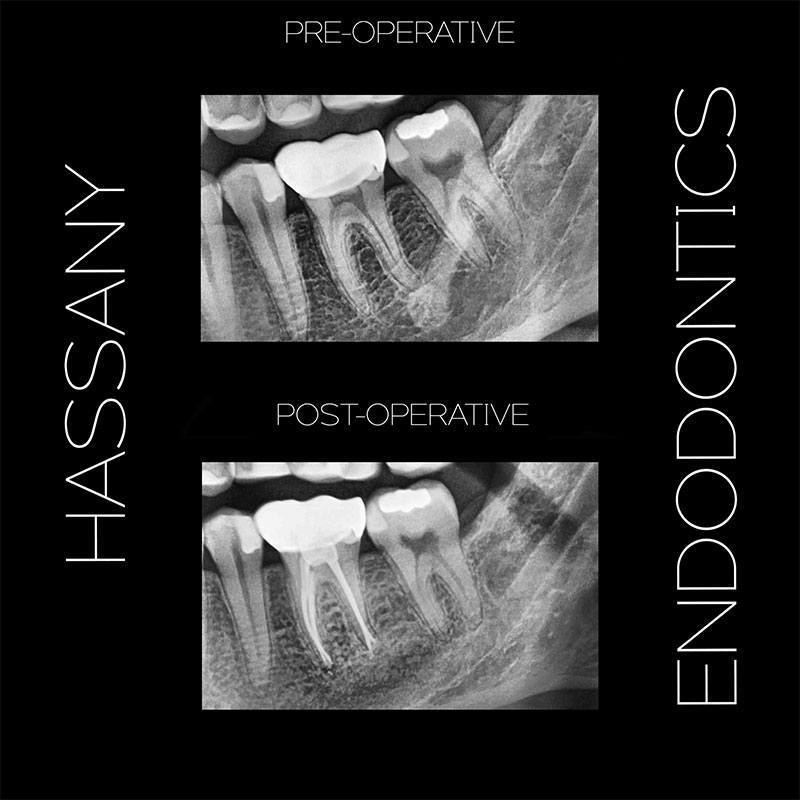
Case 2: Molar retreatment
Patient presented with chronic pain on a previously treated tooth with Sympotmatic apical periodontitis. I was skeptical of this tooth so I recommended the patient to take a CBCT to avoid any surprises. Turns out the patient had a second distal canal that as previously unfilled. Minimally invasive access cavity was done through the crown using microscope. Instrumentation was completed with Protaper Gold and Vortex Blue rotary files. Backfill technique was used for obtruation and tooth was temporized. Patient was referred to GP for final restoration.
Case 3: Endodontic surgery
Patient presented with periapical inflammation on tooth no. 30 due to separated instrument left in the periapical area during their previous endodontic treatment. Endodontic surgery was done to remove the separated instrument, curettage was done to remove periapical granulation tissue and apicoectomy was completed on both roots. Final retrograde restoration completed using EBA, flap was replaced and stutured. Patient is under observation for healing and will be referred back to their GP.
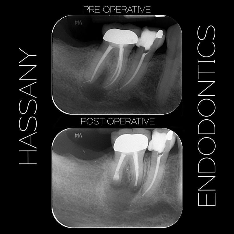
Case 4: Endodontic treatment through crown
Patient referred by GP for pain in tooth 19 upon biting, pulp testing revealed irreversible pulpitis. Patient was unable to withstand X-ray sensor and presented with limited mouth opening so we had to use limited field panoramic imaging. Access was completed through the crown, apical one third of the distal canals was very calcified but patience and size 10K files helped in provide enough patency for instrumentation with Protaper Gold files. After instrumentation was completed GP cone was used to seal the apical one third with AH26 sealer. The rest of the obturation was done with backfill technique. The entire procedure was completed in a single visit with my microscope and patient was referred back to their GP for final restoration.
Case 5: Curved canals
Patient was referred for endodontic treatment on tooth no. 17 that had a crown placed previously. Crown came off during rubber dam placement. Conservative access was done using microscope followed by extirpation and biomechanical instrumentation at working length. Rotary instrumentation was done to minimize chance of instrument separation. Obturation completed with backfill technique. Temporary filling placed and crown recemented with temporary cement. Patient was sent back to general practicioner for final restoration.
Schedule an appointment for a consultation and X-rays.


